
Meet the Team
-

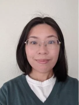
Humza Furkan Alkan
Aims to understand metabolic crosstalk between immune and melanoma cells at early stages of metastasis
-

James Amatruda
Researches pediatric sarcomas driven by fusion oncogenes. Leads the development of novel approaches to phenotypic characterization of primary pediatric cancer cells in vivo
-

Hazel Borges
Develops the clearing and labeling strategies necessary to visualize early metastatic colonization events
-

Bo-Jui Chang
Develops and applies light-sheet-based imaging techniques to live-cell and zebrafish embryo related studies
-

Bingying Chen
Develops light-sheet and multiphoton microscopes for rapid volumetric imaging with high resolution
-

Stephan Daetwyler
Develops and applies automated multi-scale microscopy to investigate the dynamic processes of metastasis in zebrafish xenografts
-

Gaudenz Danuser
Researches non-genomic mechanisms that promote plasticity in migration and proliferation/survival for cancer cells. Leads the development of high-throughput live cell imaging technologies
-

Kevin Dean
Researches and develops cutting-edge microscopy instrumentation and analysis. Leads the development of an automated, multi-scale, self-driving imaging system for early metastatic events
-

Hanieh Mazloom Farsibaf
Develops computational models to understand cancer cell signaling in the 3D morphological context
-

Reto Fiolka
Researches and develops transformative imaging technologies. Leads the development of novel imaging assays in Zebrafish xenografts to investigate metastasis mechanisms
-

Krinio Giannikou
Researches the pathogenic mechanisms of adult and pediatric cancers using cutting edge genomics, spatial and single cell -omics based technologies
-

Gabriel Muhire Gihana
Studies the morphological requirements for Ras oncogenic signaling
-

Torikul Islam
Applies light sheet microscopy and imaging techniques to detect earliest events of melanoma metastasis and study the mechanisms that determine melanoma cell fates at metastatic sites
-

Tadamoto Isogai
Researches dysregulated cytoskeleton in cancer cell survival and metastasis
-

Neha Iyer
Studies altered mechanisms related to leukemia using zebrafish & other in vitro cancer models
-

Christopher Kuo
Investigates the tumor microenvironment of Ewing sarcoma
-

Ingrid Lekk
Researches the mechanisms of pediatric sarcoma metastasis in the Zebrafish embryo
-

Jinlong Lin
Builds light-sheet microscopes capable of imaging subcellular architectures deep within tissue contexts
-

Yongfeng Luo
Develops animal models to study Ewing sarcoma pathogenesis and experimental therapeutics
-

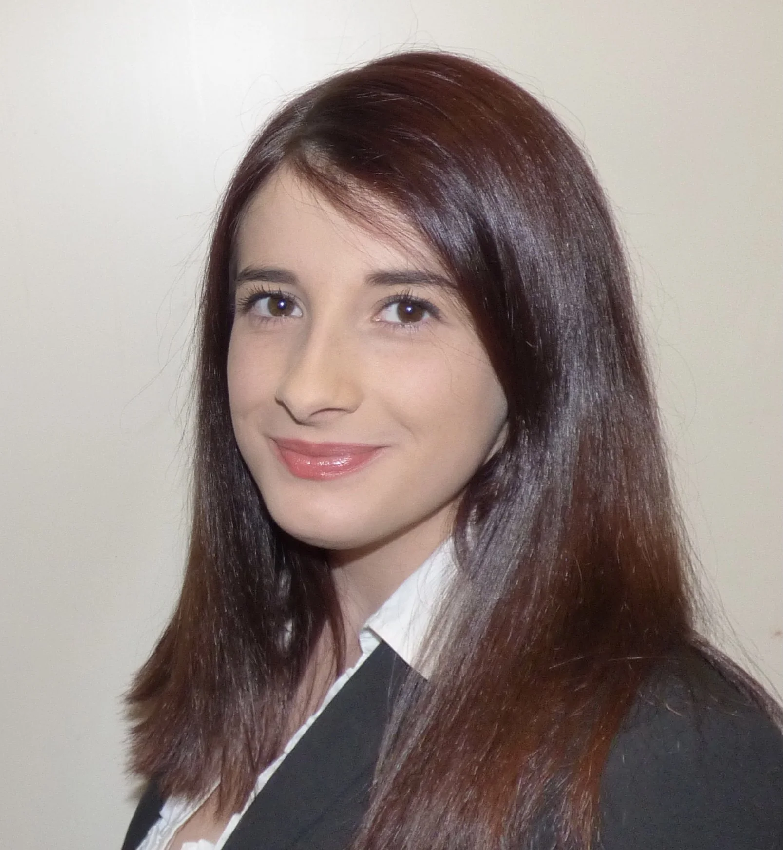
Anna Luzzi
Investigating the pathogenic mechanism of Ewing's sarcoma metastasis in Zebrafish
-

Thomas Matthews
Studies how lipid metabolism regulates oxidative stress during cancer metastasis
-

Conor McFadden
Works on developing a high-throughput zebrafish imaging system based on oblique plane microscopy to analyze cancer metastatic niches in vivo
-

Devon Moose
Studies formation of bone metastases and the effect of tumorigenesis on hematopoietic stem cells
-

Sean Morrison
Applies microscopy and imaging techniques developed by Dean and Fiolka to visualize earliest stages of melanoma metastasis for elucidation of mechanisms that determine melanoma cell fate
-

Ajit Johnson Nirmal
Studies the role of tumor microenvironment in tumor development, metastasis, and drug resistance by computational integration of high-dimensional datasets such as single-cell RNA/DNA sequencing and spatial omics
-

Shishir Pant
Studies mechanisms of therapy resistance in melanoma using highly multiplexed imaging, spatially-resolved transcriptomics, and genetically engineered tumor models
-

Divya Rajendran
Develops assays for probing the mechanism of human cancer metastasis in zebrafish embryos
-

Dagan Segal
Investigates functions of the CAV-1 gene in enabling Ewing Sarcoma metastasis
-

Zhiguo Shang
Develops algorithms & methods to address specific goals in 3D image analysis
-

Qionghua 'Nicole' Shen
Quantitative analyses of cytoskeletal alterations in early metastatic colonizing cells
-

Peter Sorger
Studies signal transduction and oncogenic mechanisms in single cells using a combination of computational and experimental methods. In this Center, the Sorger Lab leads the acquisition and analysis of highly multiplexed, single-cell imaging and genomic data
-

Elena Vasileva
Investigates the developmental origin of Ewing sarcoma using a novel zebrafish model and studies the role of proteoglycans in cancer cell transformation and tumor development
-
Xiaoding Wang
Develops autonomous microscope control software to automatically identify and interrogate rare biological phenomena
-
Manon Watzky
Studies the relationship between cell membrane changes and metastasis in Ewing sarcoma
-

Andrew Weems
Researches cellular blebbing as a morphologically encoded signaling hub, uncovering the molecular mechanisms behind this novel signaling axis and exploring its role in cancer cell survival and metastasis
-

Tao Wei
Studies how heterogeneity among melanoma cells impact melanoma cell growth and the metastatic potential
-

Clarence Yapp
Clarence has implemented deep learning image analysis methods for single cell quantification in thin tissue samples and is currently interested in applying novel 3D optical methods to characterize diseased tissue
-

Felix Zhou
Develops mathematical models of causality between cell morphology and signaling in metastatic cells
Want to join our team?
-
Dr. Amatruda is located at Children’s Hospital Los Angeles.
Drop him an email.
Check out his lab. -
Dr. Danuser is located at UT Southwestern Medical Center at Dallas.
Drop him an email.
Check out his lab. -
Dr. Dean is located at UT Southwestern Medical Center at Dallas.
Drop him an email.
Check out his lab. -
Dr. Fiolka is located at UT Southwestern Medical Center at Dallas.
Drop him an email.
Check out his lab. -
Dr. Morrison is located at the Children’s Medical Center Research Institute at UT Southwestern.
Drop him an email.
Check out his lab. -
Dr. Sorger is located at Harvard Medical School.
Drop him an email.
Check out his lab.